I want to tell the story of a beautiful phenomenon in biology. In some
sense it’s the prototype of much of the activity of life. The
phenomenon is the way in which an individual cell of
E. coli forages for nutrients. This process, known as
“chemotaxis”—the “chemo-” for chemical and the “taxis” from the Greek
τάξις, for tactics—is intelligence in one of
its most elemental forms. An individual E. coli has no brain,
obviously, and is even many orders of magnitude simpler than a human
cell, and yet already it possesses something like a sense of smell,
drive, even a memory. Chemotaxis recasts E. coli not as some
aimless gut-pest but rather as an exquisitely sophisticated
physical computer.
I’m also telling this story because
I never liked the way biology was taught in high school. It was too much about the names of things. A subject so vast is
spoiled by a textbook, which can only point at the endless parade of
stuff-there-is-to-know. It’s better approached with questions—like
“what’s happening when you smell?” or ”what is a fever,
actually?”—that contemplate narrow, deep slices.
Chemotaxis is a great slice: it’s a triumph of systems biology—we
understand it holistically but also in fine detail at almost every
level. It acquaints you with many of the most important motifs in
biology, including the way in which protein structure determines
function; how membranes control the information flow into cells; and
how chemical modifications store and communicate state. It involves
one of the most sophisticated and beautiful pieces of molecular
nanotechnology, the flagellar motor. And it helps give an intuition
for how a bag of unthinking chemicals could possibly give rise to a
being.
The 30,000-foot view
Even with simple rules, the E. coli finds food more often than not
The basic idea is this: E. coli “smells” chemicals it’s
attracted to with a set of nose-like receptors and decides how to
swim. Depending on what it senses, it can either use its flagellar
tails to swim forward—this is known as a “run”—or it can spin in a
random direction (a “tumble”). By running when the getting is good and
tumbling when it isn’t, the E. coli takes a meandering path
toward the attractant.
A little more detail now: there are half a dozen or so rotors on the
E. coli’s body, each controlling a long whip-like tail that
flows behind it. When all the rotors are spinning in the same
direction, the tails join together into a coil that torques the cell
forward into a run. When even one rotor is spinning against the
others, the coil unbundles and E. coli spins into a tumble.
In a uniform chemical environment, the E. coli swims in a
random walk by balancing runs with periodic tumbles. By default, a run
lasts about a second, or ten times longer than a tumble. The rate of
runs versus tumbles, and their relative duration, is carefully tuned
to balance “exploitation” and “exploration”: if runs happened too
often or lasted too long, E. coli would range too widely and
zip past its food; too seldom or too short, and it’d likely never find
food in the first place.
But how is this balance achieved? The crux of it is a signaling
molecule called CheY (pronounced “KEY-why”). CheY is constantly
bouncing around in the cytoplasm of the E. coli, interacting
with both the receptor complex (the “nose”) and the rotors, carrying
information between them. In the steady state, when CheY encounters
the receptor complex, it gets chemically modified, or
“phosphorylated,” at a certain rate to become CheY-p. Unlike the
unmodified version, CheY-p has a strong affinity for the rotors, and
when enough copies bind to one, it reverses its spin, causing a
tumble.
The trick is that when the nose detects an increase in the
concentration of attractant, that steady turning of CheYs into CheY-ps
is interrupted. As a result, fewer CheY-ps bind to the rotors; fewer
reversals take place; and so the E. coli runs more and
tumbles less. In other words, the all-important relative rate of runs
versus tumbles is determined entirely by how often the
CheY→CheY-p process churns—and this, in turn, is determined by
how much attractant is detected by the nose.
You can see this process in action in the interactive illustration.
Try altering the ratio of CheY-p (white) to CheY (blue) by adding some
attractant (pink). You‘ll end up inducing a long run.
Less attractant
More attractant
Why do you need all this complexity? You could imagine a system in
which the motors themselves responded directly to attractant. We’ll
see later on that the stream of CheY-ps acts as a kind of adaptable,
tunable chemical amplifier. “Bacterial cells can amplify
signals more than 50-fold; that is to say, a 2% change in receptor
occupancy can bring about a 100% change in the output of the system at
the flagellar motors. This feature allows cells to sense minute
changes in concentration—less than three molecules per cell volume!”
The story gets more complicated: adaptation
If the system were as described above, then
E. coli wouldn’t have much dynamic range. Imagine: if the
cell has a huge reaction to just three molecules of attractant,
wouldn’t a thousand times as many just completely overwhelm it?
In reality, the E. coli is able to respond sensitively across
five orders of magnitude
of attractant concentration. The cell learns to treat whatever
concentration it stumbles into as the new normal, so that the
slightest increase triggers the same hypersensitive response as
always.
The mechanism powering this adaptation is extremely clever. You can
think of each receptor as being equipped with “struts” that have
pockets in them. When the receptor is bound to attractant, its struts
change shape so that these pockets open up, and become the targets for
little molecules known as methyl groups. Methyl groups are ubiquitous
in biochemistry: for instance, they help determine which parts of your
DNA get expressed. Methyl groups bind to the structural proteins your
DNA strands coil around, called histones; the “methylated” histone can
kink the DNA strand into or out of view of your transcription
machinery, turning it on or off.
In this case, methylation serves to fill up the strut’s pockets,
causing it to become more rigid. (I’m simplifying the actual physical
details somewhat, as we’ll see later.) With more rigid struts the
receptor’s signaling power is dampened: it takes more attractant to
elicit the same response. Because there are many methylation sites per
strut and many struts per receptor, there’s a wide range of possible
dampening values—as if those pockets were really the holes of an
elaborate wind instrument.
[Bray]
This wide dynamic range is what allows the bacteria not just to find a
favorable environment but to keenly and speedily nose its way up a
chemical gradient. No wonder a similar mechanism is used by cells in
your immune system to track and hunt down invaders.
Methylation of the receptors gives E. coli a “simple chemical memory.” This is a powerful and somewhat profound idea: individual bacteria
can model their environment and remember important features of it by
encoding that information in internal chemical modifications.
E. coli “knows” whether attractant has become more or less
concentrated in its surroundings going back
several seconds; that helps it determine whether it’s swimming in a good or bad
direction. Which is not that different in principle from what brains
do. In fact one reason that it requires an artificial neural network
of about a
thousand
elements just to model the computational capabilities of a single real
neuron is that the real neuron stores so much “state” in its internal
chemistry.
(Here‘s an aside: should we be surprised at how resilient people can
be, given the mechanisms available to a single cell for accepting
previously extreme conditions as “a new normal”? No doubt our macro
resilience is in some cases actually underwritten by similar cellular
mechanisms.)
The full picture: a complex signaling network
The video above is a very legible overview of
E. coli chemotaxis, from a popular textbook. It layers in
even more detail, including not just the proteins that phosphorylate
CheY but those that dephosphorylate it; and not just the proteins that
methylate the receptors but those that demethylate it. What you come
to see is that these doers and undoers define a sort of equilibrated
circuit whose activity can be conveniently dialed up or down.
Dennis Bray describes these sorts of circuits nicely in his book,
Wetware: A Computer in Every Living Cell:
In a typical signaling pathway, proteins are continually being
modified and demodified. Kinases and phosphatases work ceaselessly
like ants in a nest, adding phosphate groups to proteins and
removing them again. It seems a pointless exercise, especially when
you consider that each cycle of addition and removal costs the cell
one molecule of ATP—one unit of precious energy. Indeed, cyclic
reactions of this kind were originally labeled “futile.” But the
adjective is misleading. The addition of phosphate groups to
proteins is the single most common reaction in cells and underpins a
large proportion of the computations they perform. Far from being
futile, this cyclic reaction provides the cell with an essential
resource: a flexible and rapidly tunable device.
If the cell really needs to change the concentration of the modified
protein very quickly, it can. All it has to do is to switch on or
shut off the phosphate-adding reaction and the concentration will
fall precipitously—at the speed of the spinning cycle. There is no
buildup of products or depletion of substrates to slow down the
process, as there would be in a linear chain of enzyme reactions.
This is a clever way to regulate the level of some protein or
metabolite. Rather than producing the thing you want via a lengthy
chain reaction, you just have this running cycle that activates and
then de-activates it, for example via phosphorylation and
de-phosphorylation. When you want more of the active version, you just
tamp down the de-activating reaction in the cycle, as if sliding down
the volume on a stereo.
Regulation in this manner via phosphorylation and dephosphorylation
(by “kinases” and “phosphatases” respectively) is an extremely general
feature of life. “About 30–50% of human proteins contain covalently
attached phosphate. [. . .] A typical mammalian cell makes use of
hundreds of distinct types of protein kinases at any moment.”
[Alberts]
In the interactive figure above, phosphorylation is represented by the
blue dots becoming white, and de-phosphorylation happens when they
turn blue again. This cycle is constantly running. The speed of the
cycle determines how quickly the cell can react to levels of
attractant. Notice that when you add some, the blue→white
reaction stops happening as much. But the blue←white reaction
keeps going at the same rate. So blue CheY proliferates, and the cell
runs more. (If the cycle spun more slowly, the blues wouldn‘t take
over so quickly.)
Down the rabbit-hole…
One thing I don’t love in presentations of chemotaxis—and of
biological concepts generally—is that they often prominently feature
flowcharts and network diagrams. In the case of chemotaxis, as you can
gather from the video above, there are many players with nearly
indistinguishable names: CheA phosphorylates CheY to become CheY-p,
and CheZ dephosphorylates it back to CheY; CheW couples CheA to the
receptors, and CheR methylates those receptors’ struts; CheB,
meanwhile, “clips off” the methyl groups added to the struts by CheR.
A network diagram is no doubt useful for organizing this sea of names
but in a sense it foregrounds the most abstract view of the process.
I’d rather try to get a sense of the parts as a living whole or in
their individual physical detail.
When you do that, it’s amazing what you find.
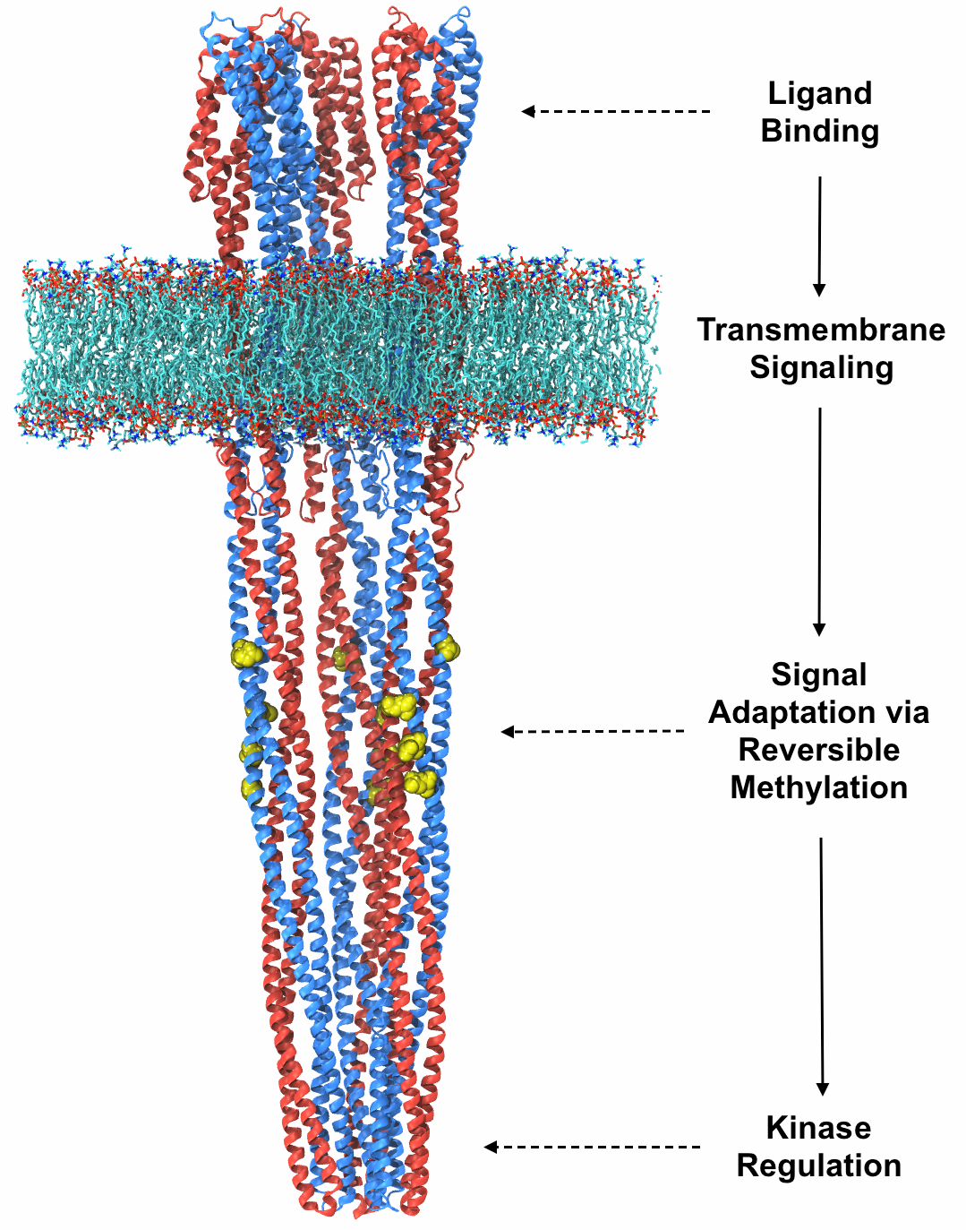
What does it mean for a receptor to detect attractant?
Almost every action in a cell depends on proteins changing shape and
binding to each other. It’s no different in the
E. coli receptor complex.
The way it works is that there are stimulus-specific proteins embedded
in the E. coli’s cell membrane, protruding into what’s known
as the periplasm. These proteins are “stimulus-specific” in the
literal sense that they are shaped so as to bind favorably with
individual molecules of attractant. E. coli has five or six
of these, for instance one that detects a crucial amino acid called
aspartate. This sensor protein has little clefts in it that are shaped
just so for molecules of aspartate to fit snugly into them.
[Falke]
In schematic form the aspartate receptor looks like this:
You can see that the sensory part—up top, where the aspartate binds—is
connected to the signaling proteins CheW and CheA by a columnar
structure that straddles the cell’s membrane. What does this protein
complex “actually” look like?
An individual protein is small enough—like a few nanometers wide—that it can’t really be seen through a regular light microscope.
This receptor from top to bottom measures about 350 angstroms, or 35
nanometers. But modern biology is all about seeing the unseeable.
Nowadays, we try to find out what nanostructures look like by
X-ray diffraction
or, more and more often, by
cryo-freezing them
in an electron microscope. Once we determine a protein’s structure
it’s usually rendered using ribbon diagrams, a style
invented by the biochemist Jane Richardson
in the late 1970s. Here’s a ribbon diagram for the
E. coli serine receptor (really it’s a “trimer of dimers,” or
a complex of six receptors):
This whole thing is the receptor. (Those parts just inside the
membrane, with little yellow methyl groups lingering stuck to them,
are the struts.) How exactly it works is quite complex, and the
subject of current research. But in simplified terms it acts like one
big piston: when the asparate binds to the part in the periplasm, the
columnar structure it’s attached to changes shape—a real biologist
would call these subtle allosteric effects; to me it looks like
dipping and tilting—in such a way to lock the thing that’s supposed to
be phosphorylating CheY, the CheA kinase, into an inactive state.
When I think of a cell I imagine a Rube Goldberg–type contraption
where an arm swings here, which drops a ball into a slide there, which
rolls down and opens a trap door, which… eventually turns on or off
some important cellular function. Indeed, E. coli’s “sense of
smell” rests ultimately in a series of physical lock-and-key
mechanisms, starting with literal molecules of e.g. aspartate nuzzling
into a protein and transmitting that physical shape-change across the
membrane.
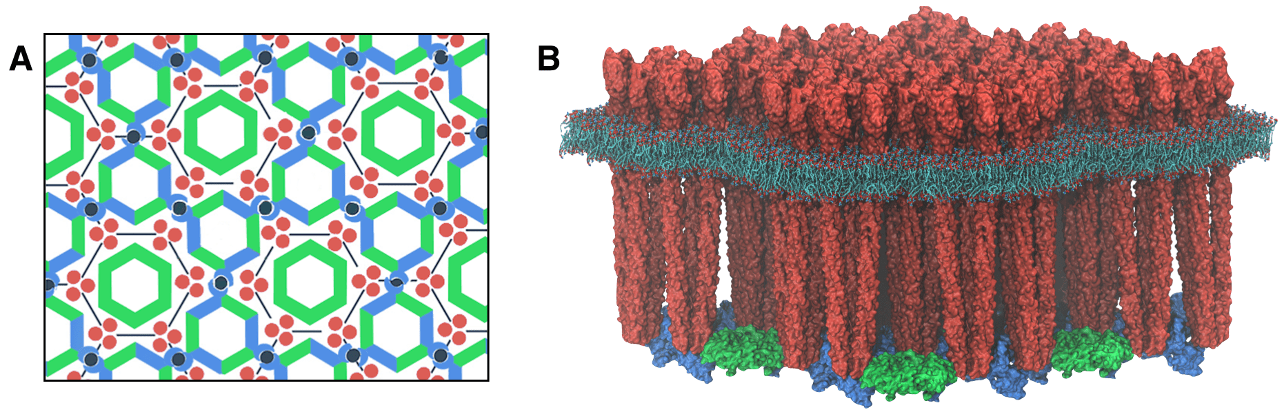
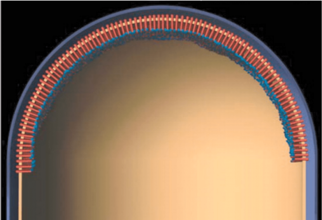
This piston-shaped receptor complex is just one of a huge array
arranged near the front of E. coli’s body. In cross section
they appear almost to have been laid down through a lithography
process, in a neat hexagonal pattern:
Calling E. coli’s receptor complex its “nose” is no mere
metaphor. Our own noses operate on a similar principle: when you smell
a flower, it means that actual flower-molecules—possibly only a tiny
number of them—have reached the inside of your nose and bound to some
protein with a specific affinity for that very molecule. This signal
is then transmitted via nerves to your brain. The human nose has
several hundred receptor proteins for smell; a dog has
more than a thousand.
Every one of our senses works like this. Touch is underwritten by
proteins that get “squished” by tactile forces into cell membranes,
triggering a set of downstream responses. Sight is my favorite
example. There’s a protein called opsin that lives in the cells of our
retina. What’s so cool about it is that the thing that changes its
shape is a literal photon. That is,
opsin converts the electromagnetic force of an incoming photon into
a biomechanical / biochemical signal. This is why I tend to think of molecular biology as the science of
shapes bumping into each other.
I think of E. coli’s receptor complex as a protoversion of
our own sensory apparatus. Its nose has only
five or six
attractant-specific sensory proteins, but their signals are
integrated, as if different sets of receptor-protein activations were
playing different “chords” on the
E. coli’s sensorium. “In short, the chemosensory array
functions as an ultrasensitive, ultrastable biological integrated
circuit or sensory chip.”
[Falke]
How the signal is carried
So a bit of attractant binds one of the receptors, and lo, the
equilibrium inside the cell begins to shift. Because the CheA kinase
is now inactive, CheYs are no longer getting phosphorylated as
quickly; the process that de-phosphorylates existing CheY-ps
starts winning out. Recall that this is a response that is dynamic, a
flow that is tuned. The net number of CheY-ps in the cell is carefully
faded down. And then what?
The CheY-ps had been binding to the flagellar rotors, flipping them,
causing tumbles. That now no longer happens as much, because the
unphosphorylated CheY doesn’t have the same affinity for the rotor as
CheY-p. As a result, the cell tumbles less, runs more, and biases its
random walk toward the attractant.
There’s something really important worth dwelling on here. When we say
that CheY-p has an “affinity” for the rotor protein, it’s not like it
gets directed there; nor does it have some long-acting magnetic
attraction for it. What this really means is that it has a strong
inclination to bind to the rotor protein when it gets really really
close to it. (And CheY, without the -p, doesn't have such an
inclination.) Given how small a single CheY-p is in the scheme of the
whole cell’s cytoplasm, it might seem improbable that it’ll somehow
sidle up right next to one of these rotor proteins somewhere on the
other end of the E. coli’s body. But that gets at the heart
of
the crazy kinetic chaos inside our cells.
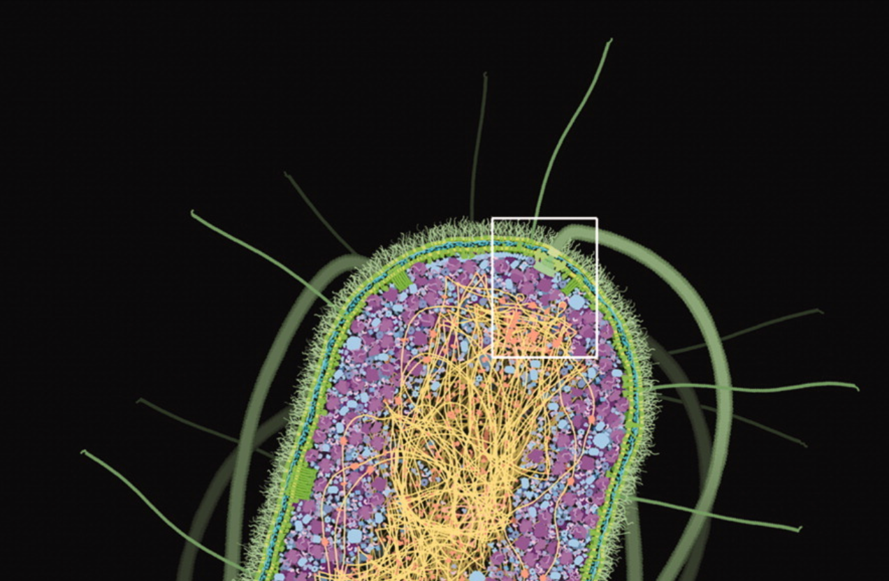
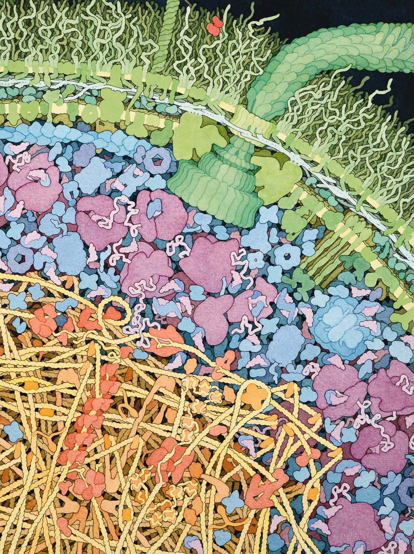
Source: David Goodsell, The Machinery of Life
Cells are dense with stuff, but everything in it is also
extremely fast-moving:
To get an idea of how fast this motion is, imagine a typical
bacterial cell, and place an enzyme at one end and a sugar molecule
at the other. They will bump around and wander through the whole
cell, encountering many molecules along the way. On average, though,
it will only take about a second for those two molecules to bump
into each other at least once. This is truly remarkable: this means
that any molecule in a typical bacterial cell, during its chaotic
journey through the cell, will encounter almost every other molecule
in a matter of seconds.
[Goodsell]
Just to put this in perspective: imagine you took an
E. coli cell and scaled it up so that it was the length of a
football field. And imagine you kept all the physics the same. A water
molecule would be about an inch wide; a protein would be about the
size of a basketball.
[BioNumbers]
The proteins would be juddering violently due to the thermal motion of
the water particles bombarding them—so violently in fact that if left
unchecked they’d be moving at 500 meters per second. But they aren’t
left unchecked: if you were in such an environment it would be so
crowded as to be nearly impossible to see. What you really get, then,
is an incredible ceaseless shaking and bouncing-into-each-other of all
the component parts.
This is why shape changes that lead to different bonding affinities
are so important in biology. It’s as if inside a cell everyone is
constantly going up to everyone else, seeing if they fit together.
Proteins sample the space of interactions with other proteins so
quickly that for a long time, most biologists didn’t really
contemplate where in the cytoplasm two reactants lived; they knew that
you never had to wait too long for them to meet each other. In fact it
was a
relatively recent discovery
that inside the cytoplasm certain proteins that share functional
relationships do seem to keep especially close together, inside little
oil drops known as “phase-separated liquids.” Weak interactive forces
between the floppy tails of different proteins cause them to
spontaneously “phase separate” into these more viscous pools, and this
biases certain proteins to interact more frequently.
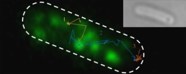
The rate-limiting step in E. coli’s reaction to attractant is
the time it takes for CheY-p to diffuse from the nose to the motor. It
takes about a tenth of a second. The journey has actually been tracked
on camera, using a fluorescent version of the protein:
Let’s talk about these motors. These things are so intricate and
beautiful and seem so reminiscent of machines we’d engineer ourselves
that they’re sometimes cited as evidence for intelligent design.
The flagellar motor operates with close to 100% energy efficiency. It
spins at about 1,500 rotations per second. And the craziest part is
that like all molecular nanomachines it is entirely self-assembled.
There’s an amazing 30-minute documentary
available on YouTube
that details the mechanics of the self-assembly process—and,
refreshingly, profiles some of the scientists who figured it out,
describing the methods they used to make their discoveries.
My favorite part of the self-assembly process is that after building a
base for the rotor, a sort of tunnel is built and the proteins that
comprise the whip-like “hook” of the flagellum are extruded through
it—as if the flagellum were built by vomiting forth parts of itself.
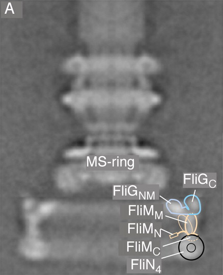
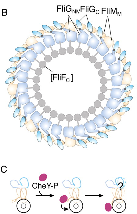
Anyway, at the base of each rotor there are a series of proteins
called FliG, FliM, and FliN—pronounced like “Fly G,” “Fly M,” “Fly
N”—to which CheY-p, our Frodo-esque bearer of the message from the
nose, attaches once it finally arrives. CheY-p has a strong affinity
for FliG and will readily glom onto it. We’ll see how that actually
affects the flagellum in a second. But for now it’s worth noting that
there’s a thresholding mechanism here: just one CheY-p attaching to
FliG won’t be enough to flip the motor from counter-clockwise to
clockwise (thereby causing a tumble)—it actually takes a handful of
CheY-ps conspiring to make that happen. In fact the motor has
something like seven states, from rotating quickly counterclockwise at
three discrete levels of decreasing speed—as if stepping through three
gears on a bike—to stalling entirely, to starting back up again in the
clockwise direction, also with three speeds.
Even as the motor is in the process of changing direction, any CheY-ps
that do attach to FliG are under constant threat of being removed by
yet another player, CheZ. That is, the proteins that would reverse the
motors are subject to removal by other proteins that un-reverse it.
Again we have a responsive regulatory circuit reminiscent of the one
that phosphorylated and de-phosphorylated CheY in the first place
upstream at the receptor. The idea is that every effect is reversible,
and in fact is reversed at a regular rate. This means that in the
absence of further signal the cell will quickly return to baseline.
How does the motor actually change directions?
As a matter of pure mechanics this might be the most ingenious part of
the story. It took quite a long time to figure out and even still it
seems that we’re not entirely confident with our explanation. But one
mechanism that’s been proposed is that CheY-p binds to a protein
called FliM (“Fly EM”) embedded in that ring that defines the base of
the rotor. This tilts it and causes a 90-degree rotation in an
attached protein called FliGc. That protein sits at the interface
between the rotating part of the motor and the so-called “stator,”
which drives it from the part that’s anchored solidly in the cell
membrane.
When FliGc changes orientation, the stepper-motor-like cycle that
normally drives the motor counterclockwise starts driving it clockwise
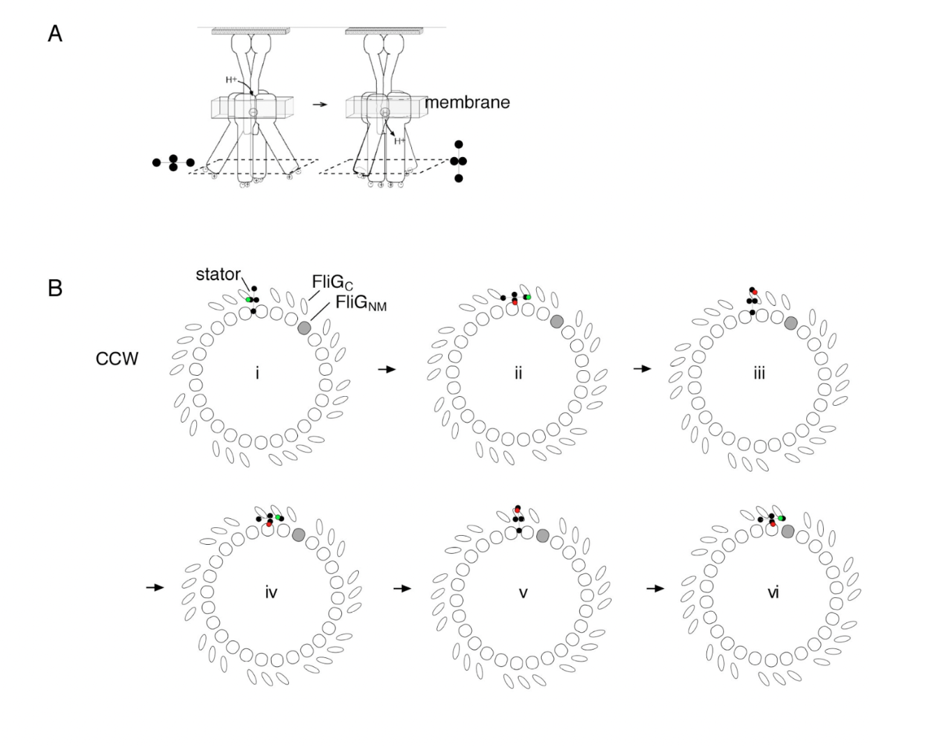
instead. In the illustration below, Figure A shows the stator, i.e.,
the driving mechanism of the motor. It works by stepping back and
forth between the “open” and “closed” states, schematized by the
and
symbols respectively. In Figure B you can see how, in the normal CCW
direction, the repeated cycling between these two states drives the
“teeth” of the motor—the crucial FliGc proteins, here tilted
left-to-right.
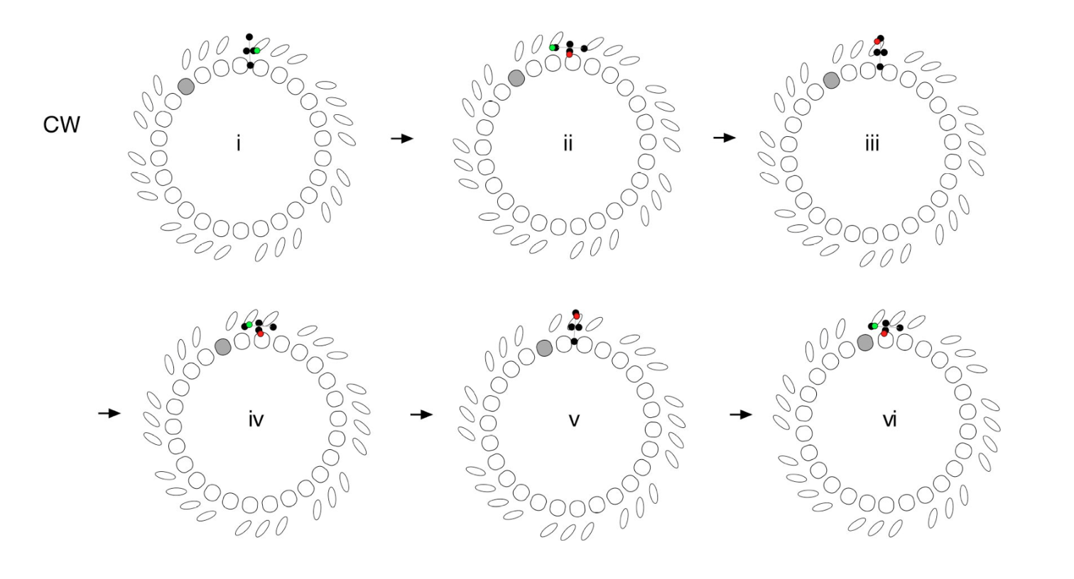
When the CheY-p arrives at the rotor it has the effect of flipping the
FliGc proteins so that now they tilt right-to-left. In that
orientation the step-drive action works the opposite way, and the
motor rotates clockwise:
You can see this more clearly below in the interactive version of
those figures. Click “Step” to drive the motor in one direction, and
“Reverse” to flip the orientation of the FliGc proteins; Step again
and you’ll see it run in the opposite direction.
Reverse
Step
It would be nice if the original paper presenting this theory included
an illustration like this. But even this crude version took me many
hours to make. As Bret Victor argues in
Stop Drawing Dead Fish, making moving pictures shouldn’t be so hard. If it were easier,
such animations would spread everywhere in scientific communication,
because so often what a paper describes is some kind of dynamic
process.
Dynamic illustrations would help readers grasp proposed mechanisms
more quickly. As it is, someone who understands a complex mechanism
usually has to explain it in patient detail to someone
else who’s good at animating; this costs time and money; and
most people simply opt not to go through with it. Perhaps someday the
process will be democratized by better tools, or by a multimodal AI
system.
How the motor changing directions causes the E. coli to
tumble
The final part of the story—for me, anyway; there’s a lot more to
explore!—is why exactly the clockwise rotation of just one of the
flagellar motors would send the whole cell a-tumbling. It helps to
understand how the thing works in “run” mode, when all the flagella
are oriented the same way.
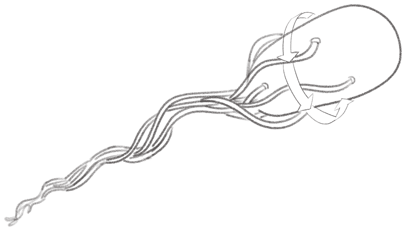
Even though this bundle of flagella sort of looks like a propeller,
when you actually think about it, that’s not really what it is. It’s
more like a pig’s curly tail that spins with a whip-y sort of motion.
How exactly does that propel the entire cell? A wonderful book called
Random Walks in Biology gets into the physics in some detail:
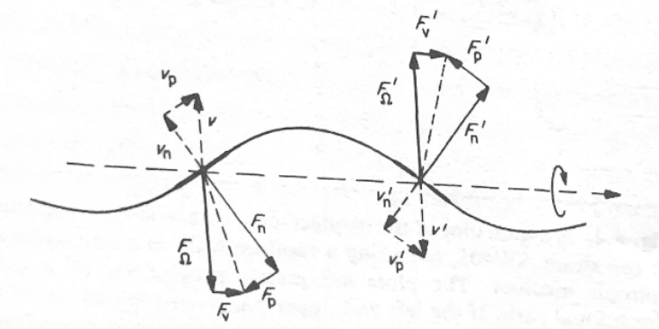
Source: Howard C. Berg, Random Walks in Biology.
“Fig. 6.3. Analysis of viscous drag on two segments of a flagellar
filament moving slowly to the right and turning rapidly
counterclockwise. The velocity of each segment, v, is
decomposed into velocities normal and parallel to the segment,
vn and vp, respectively. The segment shown on the left is moving upward in
front of the plane of the paper; the one shown on the right (denoted
by primes) is moving downward behind the plane of the paper. The
frictional drags normal and parallel to each segment,
Fn and Fp, act in directions opposite to vn and
vp, respectively. Note that their magnitudes are in the ratio
Fn/Fp = 2vn/vp. Fn and Fp are decomposed into
components normal and parallel to the helical axis,
FΩ and Fv, respectively. FΩ and F'Ω act
in opposite directions and form a couple that contributes to the
torque. Fv and F'v act in the
same direction and contribute to the thrust.”
To get a grip on things like this, it helps to have a model of some
kind that you can hold in your hand. And actually the question of how
separate filaments running in phase near each other would come to
bundle was explored nicely in
this paper. The authors used physical models of the flagella by wrapping hollow
Tygon tubes around a mandrel and filling them with epoxy. They then
used a couple stepper motors to drive the counter-clockwise rotation.
“The flow field generated by each helix tilts the other helix, causing
the helices to roll around each other and form a right-handed
wrapping”:
We tend to think of a colony of something like E. coli as an
undifferentiated evil goo, each bacterium identical to its neighbors.
But people who’ve studied these organisms under the microscope observe
a surprising amount of individual personality.
A 1976 Nature paper, “Non-genetic individuality: chance in the single cell,” explores
variation in the context of chemotaxis using strains of Salmonella and
Enterobacter bacteria.
The paper came out before the exact mechanism behind chemotactic
regulation was well-understood; all the authors knew was that “control
of tumbling can be rationalised as caused by changes in the levels of
a tumble regulator.” They hypothesized that although bacteria of the
same strain would all share the exact same DNA, there might be a
relatively small number of copies of that “tumble regulator,” and
natural variation in the transcription, translation, and destruction
of these regulator proteins could account for differences in behavior.
Their experiments were mostly at the behavioral level. They observed
how different individuals—including those in a particularly “tumbly”
mutant strain (I love that word)—reacted to environments with and
without attractant, and found plenty of variance.
Their theory was spot-on. We talked above about how
E. coli adapt to higher and higher concentrations of
attractant via a clever methylation mechanism. Well, it turns out that
the methylation of the receptor struts is governed by only about
100 CheR proteins
in the cell. The number of those proteins—along with CheB, which
un-methylates the struts—determines the speed of the “futile cycle”
that reacts to changes in attractant concentration. That is, it
affects how quickly the bacterium adapts when the concentration goes
up and refracts when it goes down.
[Gore1:06:30]
Because 100 copies of CheR is so extraordinarily tiny in the context
of the full cell buzzing with something like ten million proteins,
variation by just a handful can have a relatively large effect on the
cell’s behavior.
[Gore1:13:20]
That helps account for why different E. coli with the exact
same genetic sequence will tumble and adapt at different frequencies.
Recent
experiments
have used fluorescent microscopy to quantify the individuality of
different E. coli cells, individuality that arises not from
differences in gene expression but from the dynamics of signaling
networks.
How did we figure all this stuff out?
We don’t yet have the technology to just observe all of the activity
inside a living cell. That Goodsell painting above that shows the
crowded cytoplasm packed with proteins is an artistic composite—backed
by rigorous research to be sure—because there’s no way to capture all
the different players in situ at once. And obviously it’s a “still
life,” not a video. So how could we possibly know all this detail
about what exactly a given protein looks like, and how and when it
interacts with others to kick off some particular part of the
chemotaxis process?
There seem to be three or four major kinds of experiment. Probably the
most common is genetic: you can selectively disrupt one gene at a time
and, by observing how the mutant E. coli behaves, begin to
get a grip on each gene’s function. All of the proteins “CheY,”
“CheZ,” “CheW,” and so on are named simply because they are the
products of genes that, when excised, “cause a general defect in
chemotaxis.”
[Blair] As you
can imagine, identifying all of these is painstaking work, and
involves a considerable amount of clever inference. For instance you
might observe that without gene X the bacteria never seems to tumble;
is that because that gene is involved in recognizing attractant or in
forcing the rotor to run clockwise?
Once you have a hypothesis, a second kind of experiment involves
purifying some subset of these proteins-of-interest in vitro to see
how they work together to form a particular signaling pathway. For
example you could put CheA and CheY along with some phosphate groups
and other necessary reactants and observe whether and how much
phosphorylation takes place. That’s what the authors did in
this paper
in Cell, in 1990. They used a radioactive version of
phosphate as a tracer. “Incorporation of [32P]phosphate into CheA or
CheY was determined by excising the radioactive band out of the dried
gel and quantitating in scintillation fluid or by analysis of the
intact gel using a Phosphorimager (Molecular Dynamics, Sunnyvale, CA)
and compare with known radioactive standards.” Another common method
for observing in vitro dynamics is to genetically modify proteins to
fluoresce; or to “find” a protein in solution using an antibody that
recognizes some part of it—you attach that antibody to another
protein, and that one you fluoresce, so you can find the hidden one.
To understand the literal lock-and-key mechanics at a particular
binding site—for instance how exactly a molecule of aspartate causes a
receptor to deform, kicking off a signaling cascade—involves
“structural” biology work, i.e., taking pictures of individual
proteins or, increasingly, ensembles of them in situ. For this you can
use X-ray crystallography, nuclear magnetic resonance imaging,
cryo-electron microscopy, super-resolution light microscopy, or some
combination.
A group at
University of Illinois at Urbana-Champagne uses atomic-scale molecular
dynamics simulations, in software, to understand structural
details—like the exact
way that
CheA changes shape to kick off a downstream signaling process—that
wouldn’t be apparent from high-resolution imaging alone. (Keith
Cassidy, whose figures appeared above, now has a
lab at the University of Missouri-Columbia
that’s studying the molecular dynamics of the receptor signaling
complex.)
Sometimes you can’t get a direct picture. It may require deduction to
understand, say, how exactly a protein fits in.
One experiment
found that CheA didn’t bind to a receptor except in the presence of
CheW; that plus the fact that adding too much CheW into the mixture
actually led to a decrease in the ability of CheA–CheW complexes to
bind receptor suggested that CheW competed with that complex for the
binding site on the receptor and that therefore it must sit between
the receptor and CheW in the receptor–CheW–CheA trimer
[via Blair].
Biology is lousy with heroic inferences like that. It’s a world that’s
hard to see; sometimes you just have to imagine what’s going on down
there, and back up those imaginings with the right experiments.
The very idea that bacteria run and tumble came from experiments
published in 1972
by Howard Berg and Douglas Brown, who used a special three-dimensional
tracking microscope of their own design to watch the little suckers in
action. (A fun fact is that they called the non-runs “twiddles”
instead of “tumbles.”) Some of the physics of flagellar
propulsion—like how much force the little tails generate—was discovered
later by tethering the flagella to a microscope slide: because it’s
anchored, the “tail wags the dog“ and you can measure how fast the
E. coli’s body spins. We know that bacterial flagellar motors
are powered by the
proton motive force
from a
1977 paper
that measured how cells ran or “twiddled” in the presence or absence
of an electrical potential. But the research has become even more
refined than that. Just by observing the strength of the rotation
under various conditions—different viscosities, temperatures, and so
on—we know that “rotation is tightly coupled to proton flow, with a
fixed number of protons (~500) used to drive each revolution.”
[Blair]
Think of how detailed an understanding we’ve gotten!
One reason I’m particularly attracted to studies of
E. coli chemotaxis is that it’s an early star of what’s been
called “in silico” biology. It’s been the subject of many computer
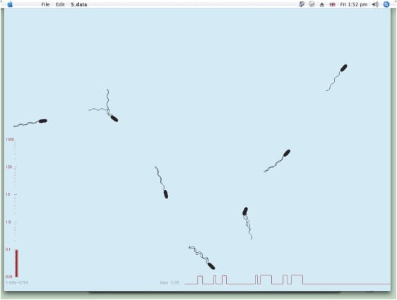
models. Dennis Bray, the author of that book that put me onto this
stuff in the first place, made one of the more
popular models. Here’s a nice screenshot of the model in action:
Maybe the chief role of a computer model is that to get it working in
the first place you have to explicitly articulate every one of your
assumptions. In much the same way that writing tends to clarify your
thinking (or at least reveal how unclear it really is), a computer
model forces you to synthesize what you know. If anything it’s even
more exacting than a blank page.
Once you have a model, you can use it to explore variations on those
assumptions. “The program gives the correct phenotype of over 60
mutants in which chemotaxis-pathway components are deleted or
overexpressed,” Bray writes. At best, a good enough model lets you
discover things you didn’t already know, or suggests your next
experiment. “In order to match the impulse response to a brief
stimulus [. . .] we also had to increase the activities of the
adaptational enzymes CheR and CheB at least an order of magnitude
greater than published values.”
So what?
Why should you care about E. coli chemotaxis? A typical
answer to that sort of question—and I’m sure the answer given in many
of the grant applications supporting the work cited here—is that there
are medical and practical uses. For instance: if you understand the
signaling pathways of bacterial chemotaxis you can disrupt them; that
work might lead to a new kind of antibiotic, which, in an era of
increasing resistance, is direly needed. Or you might hijack
chemotaxis pathways to create “intelligent sniffers” (Keith Cassidy’s
phrase) that could home in on cancer cells or environmental waste.
More generally you might say—and in fact I led with this up top—that
understanding this specific phenomenon equips you to understand all
kinds of others. “Bacterial two-component pathways
[def’n]
control a dazzling array of functions including cell division,
virulence, antibiotic resistance, metabolite fixation and utilization,
response to environmental stress, sporulation, and taxis.”
But I don’t know, to me the real reason is that it’s neat. It’s just
fun to find out about. “To learn, and at due times to repeat what one
has learnt, is that not after all a pleasure?”
 and
and
 symbols respectively. In Figure B you can see how, in the normal CCW
direction, the repeated cycling between these two states drives the
“teeth” of the motor—the crucial FliGc proteins, here tilted
left-to-right.
symbols respectively. In Figure B you can see how, in the normal CCW
direction, the repeated cycling between these two states drives the
“teeth” of the motor—the crucial FliGc proteins, here tilted
left-to-right.